39 label the transmission electron micrograph of the cell
Label This Transmission Electron Micrograph : TEM of chloroplast from ... Label the transmission electron micrograph of the nucleus. Label the transmission electron micrograph of the nucleus. Transmission electron microscopy (tem) is a microscopy technique in which a beam of electrons is transmitted through a specimen to form an image. Figures label this transmission electron micrograph ( 16, . CIN2003. Ian Roberts. medlineplus.gov › ency › articleImmune response: MedlinePlus Medical Encyclopedia Lymphocytes are a type of white blood cell. There are B and T type lymphocytes. B lymphocytes become cells that produce antibodies. Antibodies attach to a specific antigen and make it easier for the immune cells to destroy the antigen. T lymphocytes attack antigens directly and help control the immune response.
High-Pressure Freezing and Transmission Electron Microscopy to ... Transmission electron microscopy (TEM) is the main technique used to study the ultrastructure of biological samples. ... High-Pressure Freezing and Transmission Electron Microscopy to Visualize the Ultrastructure of the C. auris Cell Wall Methods Mol Biol. 2022;2517:189-201. doi: 10.1007/978-1-0716-2417-3_15. Authors Gillian ...

Label the transmission electron micrograph of the cell
Solved Label this transmission electron micrograph of | Chegg.com Question: Label this transmission electron micrograph of relaxed sarcomeres by clicking and dragging the labels to the correct location Sarcamere 1 band (light) Z disc Mline Aband (dark) H zone This problem has been solved! See the answer Show transcribed image text Expert Answer 100% (2 ratings) If you f … View the full answer Transmission Electron Microscope (TEM)- Definition, Principle, Images The working principle of the Transmission Electron Microscope (TEM) is similar to the light microscope. The major difference is that light microscopes use light rays to focus and produce an image while the TEM uses a beam of electrons to focus on the specimen, to produce an image. Electrons have a shorter wavelength in comparison to light which ... › pmc › articlesSARS-COV-2, infection, transmission, transcription ... Transmission electron micrograph of SARS-CoV-2 viral particle, isolated from a patient. Large red circles represent newly formed genome copies and the small red balloons are new virions formed by budding at the interface of Golgi apparatus and endoplasmic reticulum.
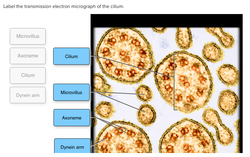
Label the transmission electron micrograph of the cell. (PDF) Label-free imaging of cell attachment with photonic crystal ... Label-free imaging of cell attachment with photonic crystal enhanced microscopy ... Bright field microscopy of HepG2/C3 cells shows cell spreading and morphology. b) Transmission intensity is plotted as a function of angle of incidence for individual pixels on (blue) and off (red) a cell. Pixel regions are highlighted in (a). ... Transmission electron micrographs showing labeling of EPS of capsular ... Download scientific diagram | Transmission electron micrographs showing labeling of EPS of capsular (b) and ropy (c-f) L. cremoris strains in reduced-fat Cheddar cheese matrix using Ricinus ... Solved Label the transmission electron micrograph of the | Chegg.com Expert Answer Answer The label is indicated from TOP to BOTTOM Ciliu … View the full answer Transcribed image text: Label the transmission electron micrograph of the cilium. Microvillus Axoneme Cilium Dynein arm Previous question Next question Labeling the Cell Flashcards | Quizlet outside the cell wall Label the transmission electron micrograph of the cell. Label the transmission electron micrograph of the mitochondrion. Label the transmission electron micrograph of the nucleus. membrane bound organelles golgi apparatus, mitochondrion, lysosome, peroxisome, rough endoplasmic reticulum nonmembrane bound organelles
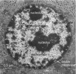
Solved Label the transmission electron micrograph of the | Chegg.com Label the transmission electron micrograph of the cell. 0 Nucleus rences Mitochondrion Heterochromatin Peroxisome Vesicle ULAR bumit Click and drag each label into the correct category to indicate whether it pertains to the cytoplasm or the plasma membrane. Label the transmission electron micrograph of the cell. 0 Nucleus ... Label the transmission electron micrograph of the cell. 0 Nucleus rences Mitochondrion Heterochromatin Peroxisome Vesicle ULAR bumit Click and drag each label into the correct category to indicate whether it pertains to the cytoplasm or the plasma membrane. › books › NBK9941Tools of Cell Biology - The Cell - NCBI Bookshelf Electron Microscopy. Because of the limited resolution of the light microscope, analysis of the details of cell structure has required the use of more powerful microscopic techniques—namely electron microscopy. Two types of electron microscopy—transmission and scanning—are widely used to study cells. In principle, transmission electron ... The Transmission Electron Microscope | CCBER Transmission electron microscopes (TEM) are microscopes that use a particle beam of electrons to visualize specimens and generate a highly-magnified image. TEMs can magnify objects up to 2 million times. In order to get a better idea of just how small that is, think of how small a cell is. It is no wonder TEMs have become so valuable within the ...
Mass Mapping of a Protein Complex with the Scanning Transmission ... A mass map of the hexagonally packed intermediate layer (HPI-layer), a regular protein monolayer from the cell envelope of Micrococcus radiodurans, has been obtained by scanning transmission electron microscopy. Samples were freeze-dried within the › cell › fulltextHomotypic fibrillization of TMEM106B across diverse ... - Cell Mar 04, 2022 · Cryo-EM and mass spectrometry-based proteomics of insoluble amyloid fibrils derived from postmortem human brains afflicted with diverse neurodegenerative diseases reveals widespread fibrillization of an endolysosomal membrane protein, TMEM106B, pointing toward a potentially pathogenic commonality between distinct proteinopathies. Transmission Electron Microscope: Definition, Parts, Working Principle ... A Transmission Electron Microscope can create a much higher resolution and magnified image than a light microscope, because of the shorter wavelength of the electron as compared to photons. ... It helps to stabilize the cells, which prevents further change or damage to the cells. This can be done in two methods; a. Chemical fixation of specimen: Lap Practical #1 EC Flashcards | Quizlet Place the following cytoplasmic structures in the appropriate structural category. Label the transmission electron micrograph of the cell. What would be the consequence if the highlighted structures suddenly became nonpolar? The lipid bilayer would not be able to hold its shape in water and the cell membrane would disassemble.
Plant Cell Diagram Electron Microscope Structure : Functions and Diagram
Tour of The Cell and Microscopy Flashcards | Quizlet Start studying Tour of The Cell and Microscopy. Learn vocabulary, terms, and more with flashcards, games, and other study tools.
Topic 1.2 Ultra-Structure of Cells - AMAZING WORLD OF SCIENCE WITH MR ...
Transmission Electron Microscope (With Diagram) The specimen to be observed is placed on a copper mesh grid. Finally, the electrons are focused by an electromagnetic projector lens (instead of an ocular lens as in a light microscope) on a screen or photographic plate. The final image in a TEM is known as transmission electron micrograph.

Electron micrograph clipart - Clipground
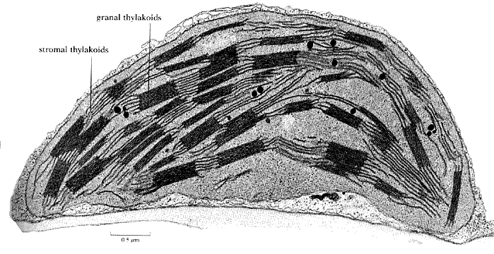
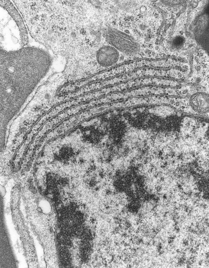
PDF Identifying Organelles from an Electron Micrograph The photograph shown below details chloroplast structure as viewed with a transmission electron microscope Courtesy of Dr. Julian Thorpe - EM & FACS Lab, Biological Sciences University Of Sussex A single Granum Chloroplast envelope visible as two membranes Stroma containing numerous small ribosomes Lamellae connecting different grana
Tem Of Endoplasmic Reticulum In Mammalian Cell Photograph by Nibsc
Study Chapter 14 & 15 Flashcards Flashcards | Quizlet Key concepts: Physical Barriers Of Innate Immunity Terminal Bronchioles Divide Into Mast Cells And Basophils Terms in this set (101) B-cells may be stimulated to transform into memory cells. True Viruses and self-proteins are examples of proteins produced inside of the cell. True Foreign antigens presented on class I MHC molecules...
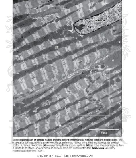
Electron Micrograph of Cardiac Muscle Showing Salient Ultrastructural ...
Label the transmission electron micrograph of the nucleus. FIGURE 2.5 Cells as seen in a transmission electron microscope. (a) Electron micrograph of a plant cell with chloroplasts in the cytoplasm around the large central vacuole. ... Label the transmission electron micrograph of the cell. 0 Nucleus rences Mitochondrion Heterochromatin Peroxisome Vesicle ULAR bumit Click and drag each label into the ...
Solved: Label The Transmission Electron Micrograph Of The ... | Chegg.com
AP Unit 2 Exam Flashcards | Quizlet Label the transmission electron micrograph based on the hints provided. Plasma cells produce antibody molecules. Name the cells included in the mononuclear phagocytic system. macrophages monocytes neutrophils An immunoglobulin molecule is an antigen secreted by T lymphocytes. False trabecula of spleen thoracic duct Disease-causing agents are called
Cells | The A Level Biologist - Your Hub
Transmission electron micrograph of U20S cells (whole mount) after ... Download scientific diagram | Transmission electron micrograph of U20S cells (whole mount) after labeling with mAb J143 and goat anti-mouse IgG coupled to 40/zm gold. The rectangular frame (frame ...

Pancreatic acinar cell, TEM - Stock Image - C015/3279 - Science Photo ...
en.wikipedia.org › wiki › Electron_microscopeElectron microscope - Wikipedia An electron microscope is a microscope that uses a beam of accelerated electrons as a source of illumination. As the wavelength of an electron can be up to 100,000 times shorter than that of visible light photons, electron microscopes have a higher resolving power than light microscopes and can reveal the structure of smaller objects.

Cell Encyclopedia: 2.2.3 Identify structures from 2.2.1 in electron ...
Electron Micrographs** Also, be sure to observe any electron micrographs which are made available in the laboratory by the instructor. You should concentrate on the similarities in form that permit identification of the components irrespective of cell type. Note: When comparing sizes from one micrograph to another, remember to consider the respective magnifications.
Post a Comment for "39 label the transmission electron micrograph of the cell"