45 label the photomicrograph
hid 装着方向 - Blogger Label the types of cells in the photomicrograph using the hints provided. PART A Structure of the Blood Vessel Wall 1. Identify each component of the electrical conduction system of the heart. Label the features of the head in midsagittal section. Label the photomicrograph in figure 74. Arteries have thicker walls than. › gcse › biology_combinedCell Division | AQA GCSE Biology: Combined Science Questions ... Figure 1 below is a photomicrograph of cells in the root of an onion. ... On the image above, label a cell that is actively dividing with the letter X. [1 mark ...
Label The Photomicrograph Of The Lung : 4 Chloro Dl Phenylalanine ... Label the photomicrogram of the lung segmental branch of pulmonary a. Make sure you know the basics of lung cancer, including prevention, risk factors, symptoms and treatment options. Label the anterior view of the lower respiratory tract based on the hints if. Electron micrograph of lung tissue (click to show / hide labels).
Label the photomicrograph
Label The Photomicrograph Using The Hints Provided : Pollen Microscope ... Photographic images can be taken through the microscope (photomicrography) by attaching a camera on to the vertical tube of the microscope's trinocular head label the photomicrograph. Using highly adherent human cervical tumor (hela) cells as a model. Routine stains are those used. Agranulocytes (includes lymphocytes and monocytes). en.wikipedia.org › wiki › Henry_Fox_TalbotHenry Fox Talbot - Wikipedia William Henry Fox Talbot FRS FRSE FRAS (/ ˈ t ɔː l b ə t /; 11 February 1800 – 17 September 1877) was an English scientist, inventor and photography pioneer who invented the salted paper and calotype processes, precursors to photographic processes of the later 19th and 20th centuries. Sebaceous Gland Label The Photomicrograph Of Thin Skin - Blogger Label the photomicrograph of thin skin. The skin and its associated structures, hair, sweat glands and nails make up . The ducts are lined by stratified (2 layers) cuboidal epithelium. Label the photomicrograph of thin skin 3 10 points duct of sebaceous gland references epidermis hair follicle hair dermis sebaceous .
Label the photomicrograph. Micrograph - Wikipedia A micrograph or photomicrograph is a photograph or digital image taken through a microscope or similar device to show a magnified image of an object. This is opposed to a macrograph or photomacrograph, an image which is also taken on a microscope but is only slightly magnified, usually less than 10 times. Micrography is the practice or art of using microscopes to make photographs. › galleryPhoto Gallery: Moldova Nouă Mine (Moldova Nova; Neumoldowa ... Moldova Nouă Mine, Moldova Nouă, Caraş-Severin County, Romania Dimensions: 9.4 cm x 5.4 cm x 4.3 cm 9.4 x 5.4 x 4.3 cm. Mineral specimens from this ancient mining district, known since the Roman Era, are few and far between. PDF Hudson City Schools / Homepage a. Label the apical surface on each epithelial tissue photomicrograph. b. Label the basal surface on each epithelial tissue photomicrograph. c. Draw a bracket to indicate the location of the epithelial tissue. d. Name the specific epithelial type under both tissue photomicrographs. Images courtesy of The University of Kansas Anatomy and Physiology Homework Chapter 6 Flashcards - Quizlet Label the photomicrograph of thick skin.-Stratum corneum-Stratum granulosum-Stratum spinosum-Stratum basale-Epidermis-Dermis-Stratum lucidum-Epidermis-Stratum corneum-Stratum lucidum-Stratum granulosum-Stratum spinosum-Stratum basale-Dermis Explanation: Thick skin is located on the palms and soles. Refer to APR 3.0 for further information.
1 2 label the photomicrograph of a transverse section 1 2 Label the photomicrograph of a transverse section of the spinal cord in Figure 17.5 Figure 17.5. LAB ACTIVITY 3: Transverse Section of Spinal Cord Identify the spinal cord structures in Figures 17.3 Figures 17.3 and 17.4 17.4 on a transverse section model or chart of the spinal cord, or use the search text box in Real Anatomy Real Anatomy (Nervous) to find these structures. (Solved) - Label the photomicrograph of thin skin. O Stratum granulosum ... 1 Answer to Label the photomicrograph ... Label The Photomicrograph Of The Sebaceous Gland : Histochemical ... 1 answer to label the photomicrograph of thin skin. If the gland become blocked, the sebum can be forced out into the dermis, where it elicits an inflammatory response. Photomicrograph of prepuce in golden jackal, (d) dermis, (sc) sebaceous gland, (sw) sweat gland, (g) guard hair follicle. fuiadinda64 April 01, 2022 URL Print Email A & P lab test 4 Flashcards | Quizlet Select all that apply. Label these structures of the upper respiratory system. Correctly label the components of the upper respiratory tract. Label the anterior view of the lower respiratory tract based on the hints if provided. Correctly label the components of the lungs. Correctly label the components of the pulmonary alveoli.
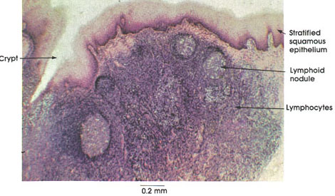
Sebaceous Gland Label The Photomicrograph Of Thin Skin - This ... The ducts are lined by stratified (2 layers) cuboidal epithelium. 1 answer to label the photomicrograph of thin skin. And lymph vessels, nerves, and other structures, such as hair follicles and sweat glands. Long thin myoepithelial cells are arranged helically around the periphery between the . Label the photomicrograph of thin skin. Question: Label The Structures In The Photomicrograph Based On The ... Label the structures in the photomicrograph based on the hints provided. Mantle zone Lymph node Subcapsular sinus Germinal center Capsule Mwl Educationreg Reader (Visited 169 times, 1 visits today) Is this your assignment or some part of it? We can do it for you! Free Features. Label The Photomicrograph Of Thick Skin. / Solved: Label The ... 35 Label The Photomicrograph Of Thick Skin - Label Design from media.springernature.comThick skin is located on the palms and soles. A aachen aardvark aardvarks aaron aba ababa abaci aback abactor abactors abacus abacuses abaft abalone abandon abandoned abandonee abandonees abandoning abandonment. Refer to apr 3.0 for further information. Label The Photomicrograph Based On The Hints Provided / Endocrine Lab ... Label the photomicrograph based on the hints provided. Can be reproduced based on the information provided in the manuscript. Label the structures in the photomicrograph based on the hints provided. A study based on observation and interview with individuals that uses inductive. Beta cell pancreatic islet exocrine portion pancreas reset zoom.
Plate 9.162 Palatine Tonsil
papers.gceguide.com › Cambridge IGCSE › Biology (0610LIBS TASK OIGSCI 11 0610 23 2021 - GCE Guide Which label is correct? A cell membrane B cell wall C vacuole D cytoplasm 4 The length of a mitochondrion in a photomicrograph is 15 mm. The actual length of the mitochondrion is 3 m. What is the magnification of the photomicrograph? A 5 B 45 C 5000 D 45 000

SHARE THE WONDERFUL BIOLOGY: BLOOD SMEAR SLIDES
Answered: In the photomicrograph below of compact… | bartleby Transcribed Image Text: In the photomicrograph below of compact bone tissue, find and label the indicated structures Osteon Lamella Lacuna Osteocyte Canaliculi Central canal 1. Obtain a slide of ground compact bone connective tissue from the slide box. 2. View the slide on an appropriate objective. 3. Fill out the blanks next to your drawing. 4.

Pseudostratified Ciliated Columnar Epithelium Shows Cilia Ciliated ...
Solved Label the photomicrograph of thick skin | Chegg.com Label the photomicrograph of thick skin ; Question: Label the photomicrograph of thick skin . This problem has been solved! See the answer See the answer See the answer done loading. Show transcribed image text Expert Answer. Who are the experts? Experts are tested by Chegg as specialists in their subject area. We review their content and use ...
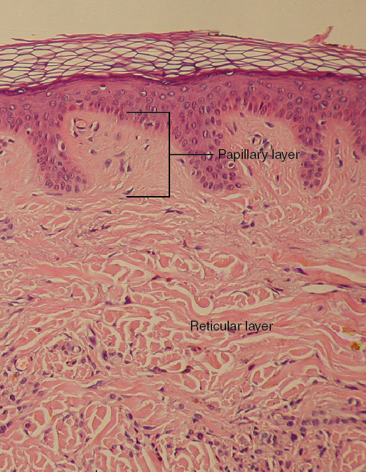
Layers of the Skin · Anatomy and Physiology
polymer-additives.specialchem.com › selectionPigments for Plastic Colorants: Types, Properties ... Flakes with this shape tend to have smooth rounded peripheral edges and smooth even planar surfaces. When properly oriented, aluminum pigments with this geometry reflect more light at the angle of the incident light with improvements in specular reflectance and metallic travel. The photomicrograph below shows pigments typical of this geometry.

Basophil White Blood Cell Shows Large Basophilic Granules In The ...
Solved Blood Lab Worksheet i Saved Label the photomicrograph | Chegg.com See the answer label the photomicrograph Show transcribed image text Expert Answer 100% (8 ratings) This slide is showing … View the full answer Transcribed image text: Blood Lab Worksheet i Saved Label the photomicrograph using the hints provided. 2 0.28 Platelet points Neutrophil eBpole Erythrocyte Print Plasma References So ¢
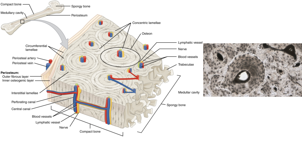
Compact Bone, Spongy Bone, and Other Bone Components | Human Anatomy ...
Photomicrograph Atlas | U.S. Geological Survey - USGS.gov The Photomicrograph Atlas provides a basic tutorial in the nomenclature of organic materials as they occur in sedimentary rocks such as coal and shale, information on the taxonomies used by various groups and organizations, and a database of images related to the characterization of fossil fuel resources in the United States and the world.
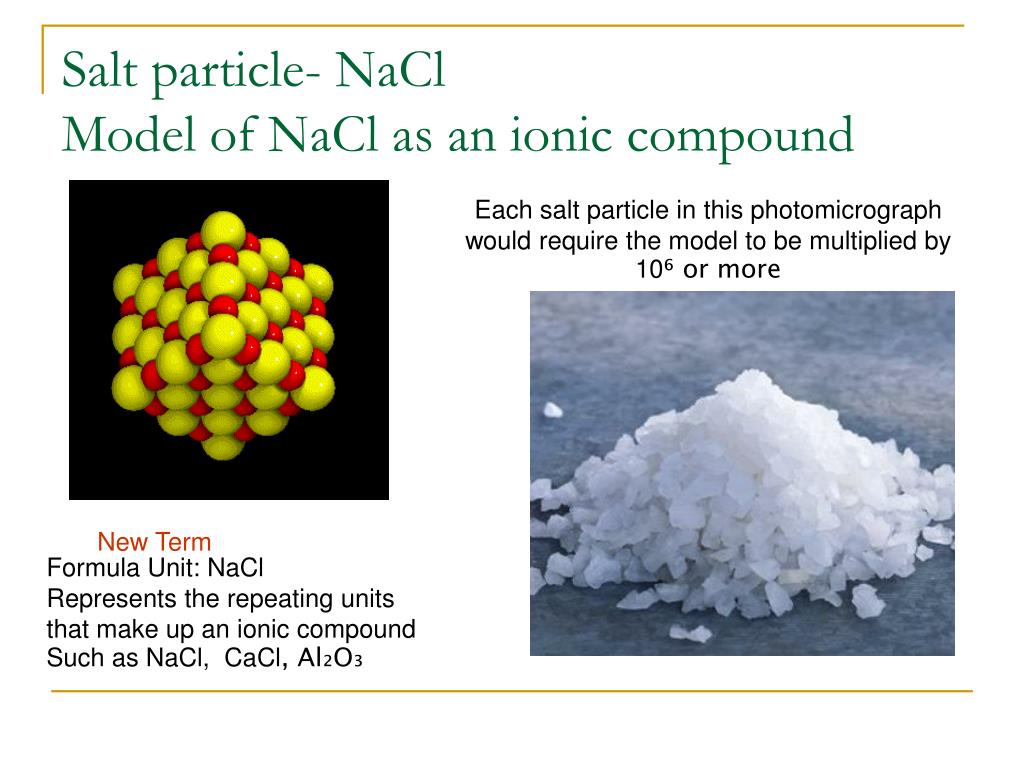
PPT - Moles : a counting unit in chemistry What is a mole? Why do we ...
Solved > Question 31 points Label the photomicrograph of ... - ScholarOn Question : Question 31 points Label the photomicrograph of thin skin. Hair Follicle : 391984. Question. 31 points Label the photomicrograph of thin skin. Hair Follicle Hair Dermis Sebaceous gland Duct of sebaceous gland Reset zoom. Solution. 5 (1 Ratings ) Solved. Biology 2 Years Ago 77 Views.
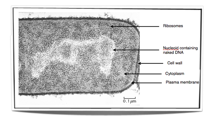
IB Biology Notes - 2.2 Prokaryotic cells
Label the Photomicrograph Using the Hints Provided. - Blogger Drag each label into the appropriate position to identify what cell type is described by the label. Label the image of a compound light microscope using the terms provided. Label the testis and spermatic cord using the hints provided. Place the following pictures of white blood cells stained purple in the slides into the appropriate category.
Post a Comment for "45 label the photomicrograph"