38 label microscope diagram
Labelled diagram - Wordwall Labelled diagram Drag and drop the pins to their correct place on the image. Examples. What are they doing? by Nataliapisettas. Labelled diagram. Sequencing Session 22. by Jkanapes1. Labelled diagram. Label Fractions on Number Line. by Sarahbutler. Labelled diagram. Female Reproductive System. by Hickskel. Labelled diagram. 8.1 Label the sentence. by Christianjolene. Labelled … Ternary Phase Diagram - an overview | ScienceDirect Topics Ternary phase diagrams are used to represent all possible mixtures of three solvents [1]; they are described in Chapter 3.Here, we shall indicate how they should be used to minimize the solvent consumption. Figure 2.1 (top) shows the methanol–chloroform–water ternary phase diagram with the tie-lines in the biphasic domain. Five particular compositions are shown in the …
Virtual Labs: Using the Microscope - GameUp - BrainPOP. In this free online science interactive, students learn the procedures for operating a compound optical light microscope as they would use in a science lab. bVX0-zncj9qJ3G1_r18rkIpQL02X-Oi6tWViR4g4-vwDVmU50WZA-4bRZMjM2TXmc88PAkJ1g0jIembnEbM

Label microscope diagram
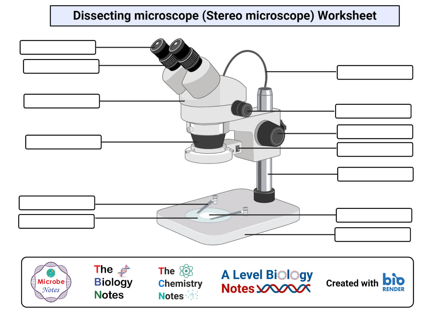
Welcome to Virtual Urchin - University of Washington microscope compare. specimen compare. development & embryology. fertilization lab. embryogenesis to hatching. analyzing gene function. ecology & environment. our acidifying ocean. predator & prey. surfing to settlement. basic biology. urchin anatomy. about us. teacher resources. useful links. Select Language: Welcome to the new Virtual Urchin website! Major … Parts of Stereo Microscope (Dissecting microscope) – labeled diagram ... Labeled part diagram of a stereo microscope Major structural parts of a stereo microscope. There are three major structural parts of a stereo microscope. The viewing Head includes the upper part of the microscope, which houses the most critical optical components, including the eyepiece, objective lens, and light source of the microscope. Electron microscope - Wikipedia An electron microscope is a microscope that uses a beam of accelerated electrons as a source of illumination. As the wavelength of an electron can be up to 100,000 times shorter than that of visible light photons, electron microscopes have a higher resolving power than light microscopes and can reveal the structure of smaller objects.. Electron microscopes use shaped magnetic …
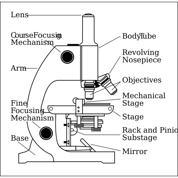
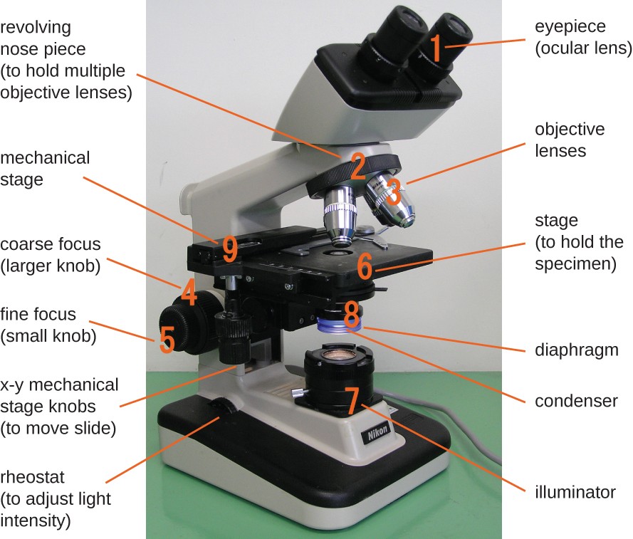
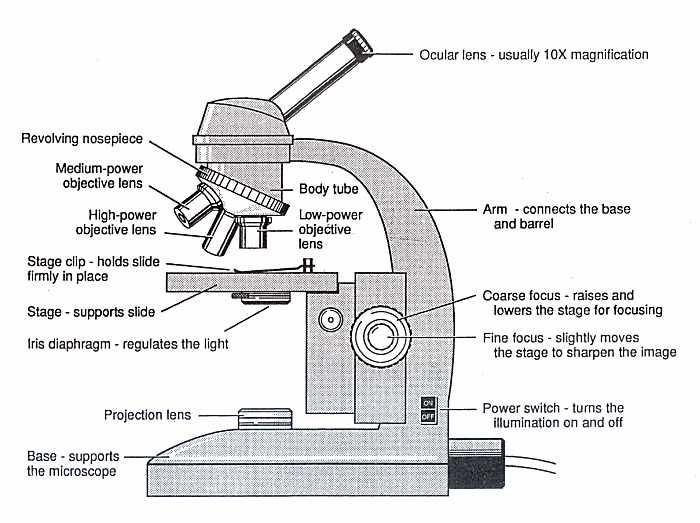
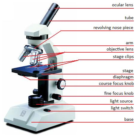
Label microscope diagram. Microscope, Microscope Parts, Labeled Diagram, and Functions 19.01.2022 · Revolving Nosepiece or Turret: Turret is the part of the microscope that holds two or multiple objective lenses and helps to rotate objective lenses and also helps to easily change power. Objective Lenses: Three are 3 or 4 objective lenses on a microscope. The objective lenses almost always consist of 4x, 10x, 40x and 100x powers. The most common eyepiece lens is … Label the microscope — Science Learning Hub 08.06.2018 · All microscopes share features in common. In this interactive, you can label the different parts of a microscope. Use this with the Microscope parts activity to help students identify and label the main parts of a microscope and then describe their functions.. Drag and drop the text labels onto the microscope diagram. If you want to redo an answer, click on the … Parts of a microscope with functions and labeled diagram Apr 19, 2022 · Figure: Diagram of parts of a microscope. There are three structural parts of the microscope i.e. head, base, and arm. Head – This is also known as the body. It carries the optical parts in the upper part of the microscope. Base – It acts as microscopes support. It also carries microscopic illuminators. Fluorescence microscope - Wikipedia The majority of fluorescence microscopes, especially those used in the life sciences, are of the epifluorescence design shown in the diagram.Light of the excitation wavelength illuminates the specimen through the objective lens. The fluorescence emitted by the specimen is focused to the detector by the same objective that is used for the excitation which for greater resolution will …
Electron microscope - Wikipedia An electron microscope is a microscope that uses a beam of accelerated electrons as a source of illumination. As the wavelength of an electron can be up to 100,000 times shorter than that of visible light photons, electron microscopes have a higher resolving power than light microscopes and can reveal the structure of smaller objects.. Electron microscopes use shaped magnetic … Parts of Stereo Microscope (Dissecting microscope) – labeled diagram ... Labeled part diagram of a stereo microscope Major structural parts of a stereo microscope. There are three major structural parts of a stereo microscope. The viewing Head includes the upper part of the microscope, which houses the most critical optical components, including the eyepiece, objective lens, and light source of the microscope. Welcome to Virtual Urchin - University of Washington microscope compare. specimen compare. development & embryology. fertilization lab. embryogenesis to hatching. analyzing gene function. ecology & environment. our acidifying ocean. predator & prey. surfing to settlement. basic biology. urchin anatomy. about us. teacher resources. useful links. Select Language: Welcome to the new Virtual Urchin website! Major …
Parts of a microscope with functions and labeled diagram

Simple Microscope- Definition, Principle, Magnification ...
Parts of a microscope with functions and labeled diagram
Label a microscope - Teaching resources

Label Microscope Diagram | Microscope parts, Microscope ...
Name Date Sci STANDARD MICROSCOPE DIAGRAM Label only the ...

Diagram of diatom microscope slide positioned with its label ...
What is a diagram of a plant and animal cell under an ...

Biology label part of microscope

Parts of a Microscope Labeling Activity

Label the Microscope Diagram | Quizlet
Microscope Biology - 2022

Microscope parts 3D learning APK for Android Download

Label the microscope — Science Learning Hub
Instruments of Microscopy | Microbiology | | Course Hero

Cytology. Cytology. radiation used to illuminate the specimen ...
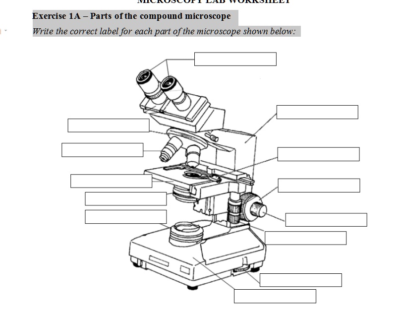
SOLVED: Exercise 1A _ Parts ofthe compound microscope Write ...
Free Microscope Drawing, Download Free Microscope Drawing png ...
Label a microscope - Teaching resources
Simple Microscope - Parts, Functions, Diagram and Labelling ...

Label the light microscope | Teaching Resources

Microscope Diagram Labeled, Unlabeled and Blank | Parts of a ...
Label Microscope Parts - ClipArt Best
Microscope Parts and Function

Parts of a Microscope with Their Functions • Microbe Online

File:Labelledmicroscope.gif - Wikimedia Commons
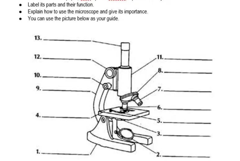
Answered: Label its parts and their function.… | bartleby

This is a common compound microscope. Label its parts from A ...

Compound Microscope Parts, Diagram Definition, Application ...

Label the diagram of the microscope and explain the role of ...
Lab - Microscope: MAH-Summer 2019-Anatomy and Physiology I

Compound Microscope: Parts of Compound Microscope

Label the Microscope Diagram | Download Scientific Diagram

PSB Microscopy Worksheet - Microscopy Worksheet In order to ...

The Microscope
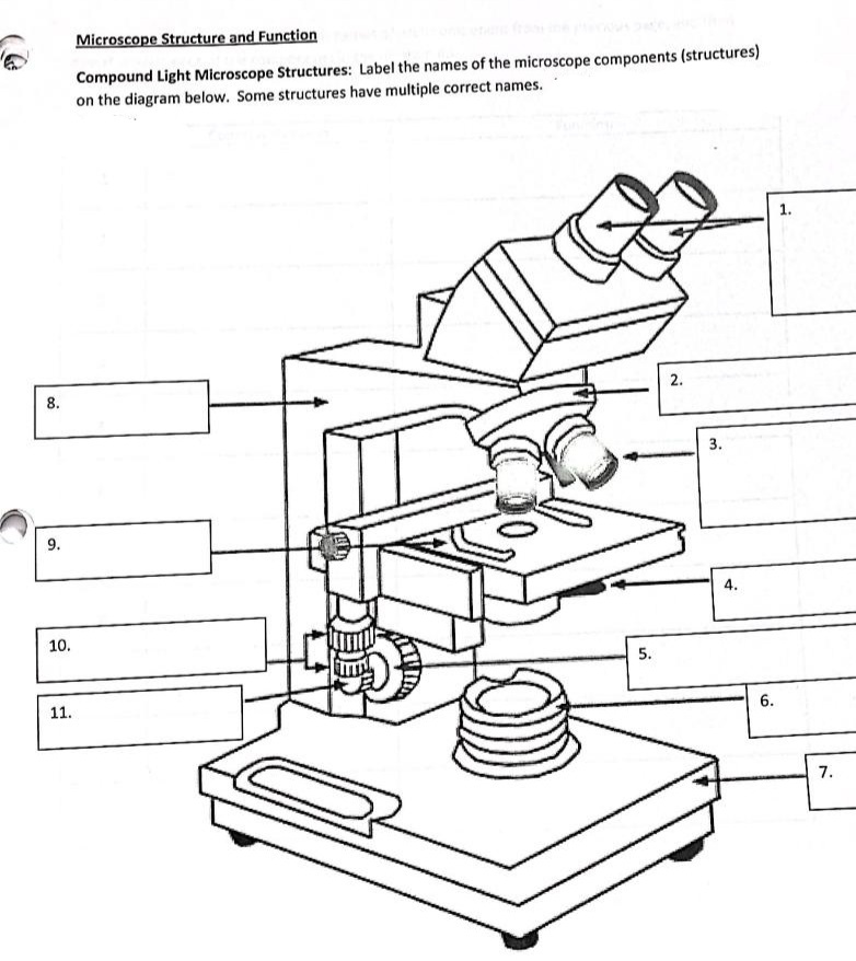
Answered: Microscope Structure and Function… | bartleby

ABOUT MICROSCOPES | Scienceart

How to draw compound of Microscope easily - step by step
Post a Comment for "38 label microscope diagram"