42 draw and label microscope
How To Draw Microscope Images: A Step By Step Guide Once you have adjusted the microscope and gathered the necessary tools, you can begin to draw what you see under the microscope. Start with a rough sketch: Begin by making a rough sketch of the specimen, indicating the overall shape and size. Observe the details: Look closely at the specimen and observe the details. Take note of the textures, colors, and patterns. 1.5: Microscopy - Biology LibreTexts Gently scrape the inside of your cheek with a toothpick and swirl it in the dye on the slide. Place a cover slip on the suspension and view at 1000X total magnification. Draw 1-3 cells large enough to show the detail that you see in your lab manual. Label its cell membrane, cytoplasm and nucleus.
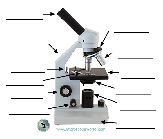
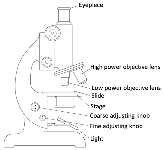
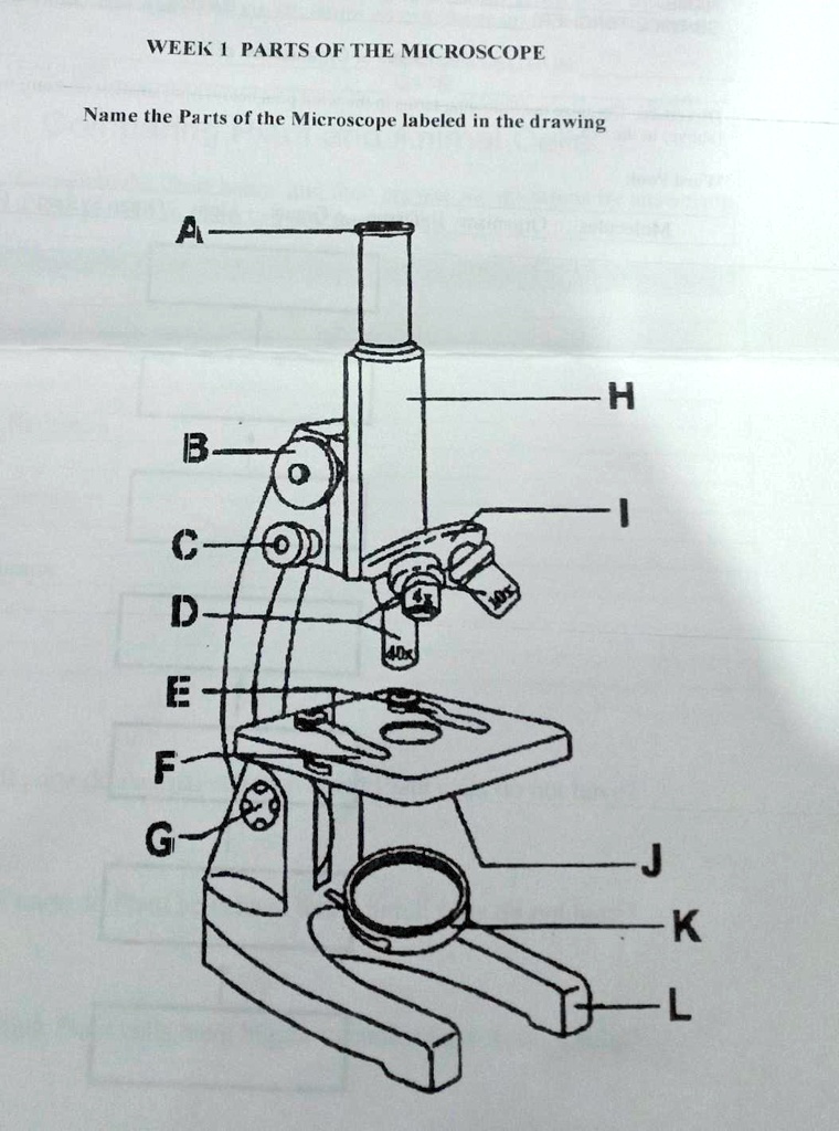
Label the microscope — Science Learning Hub In this interactive, you can label the different parts of a microscope. Use this with the Microscope parts activity to help students identify and label the main parts of a microscope and then describe their functions. Drag and drop the text labels onto the microscope diagram. If you want to redo an answer, click on the box and the answer will ...

Draw and label microscope
Label a Microscope - Storyboard That Create a poster that labels the parts of a microscope and includes descriptions of what each part does. Click "Start Assignment". Use a landscape poster layout (large or small). Search for a diagram of a microscope. Using arrows and textables label each part of the microscope and describe its function. Copy This Storyboard. How To Draw A Microscope 🔬 - YouTube #microscope #howtodraw #adimushowThis is an easy and simple drawing of microscope .This will teach you how to draw and label a microscope .This is a step-by-... Microscopy: Intro to microscopes & how they work (article) - Khan Academy Magnification is a measure of how much larger a microscope (or set of lenses within a microscope) causes an object to appear. For instance, the light microscopes typically used in high schools and colleges magnify up to about 400 times actual size. So, something that was 1 mm wide in real life would be 400 mm wide in the microscope image.
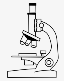
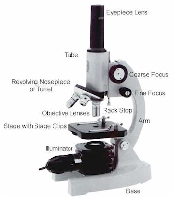
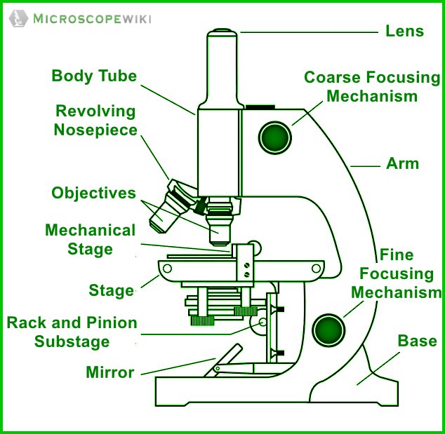
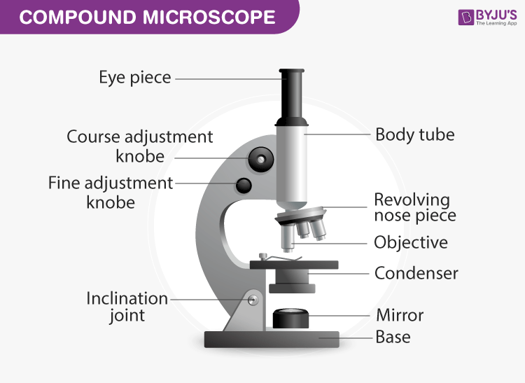
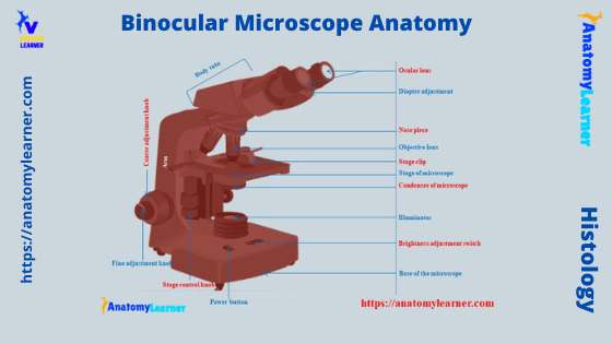
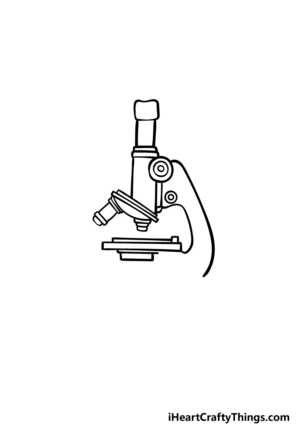
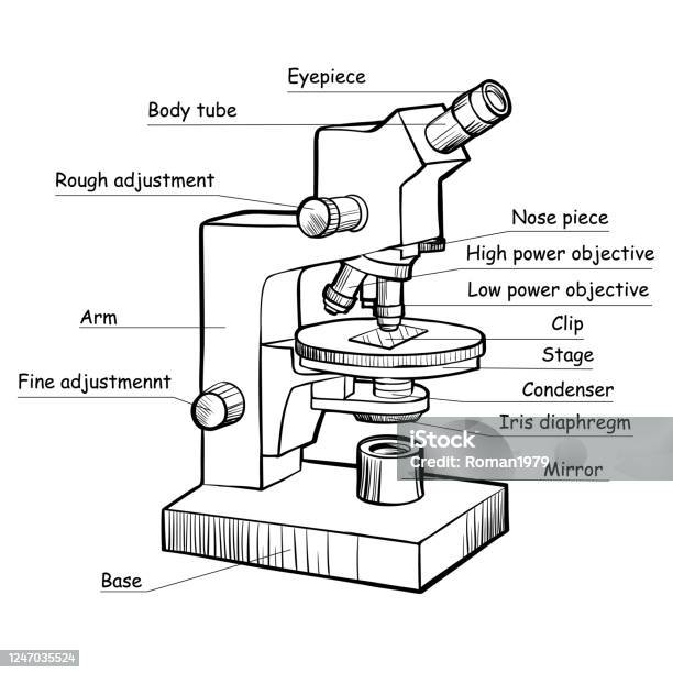
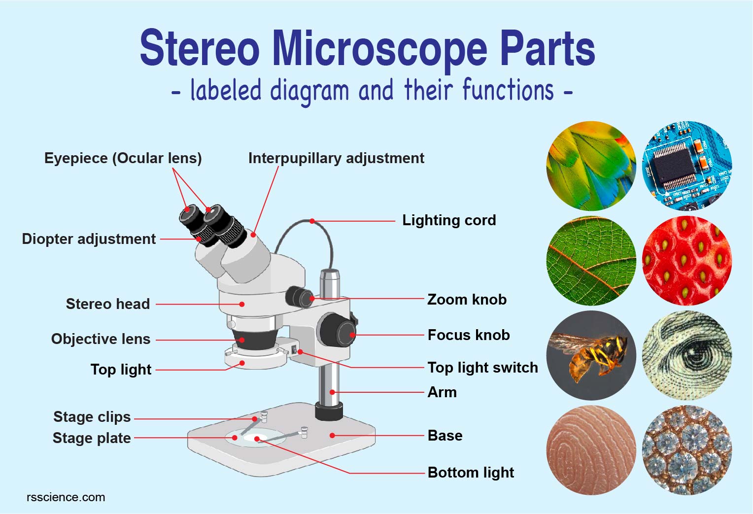
Draw and label microscope. How to draw Microscope diagram for beginners - step by step Today I will show you " How to draw Microscope diagram for beginners - step by step". Every week new drawing. Continue follow my channel and like, share,comm... Microscope Parts and Functions Eyepiece: The lens the viewer looks through to see the specimen. The eyepiece usually contains a 10X or 15X power lens. Diopter Adjustment: Useful as a means to change focus on one eyepiece so as to correct for any difference in vision between your two eyes. Body tube (Head): The body tube connects the eyepiece to the objective lenses. Arm: The arm connects the body tube to the base of the ... How to Draw a Microscope - Really Easy Drawing Tutorial 3. Use a curved line to enclose a rounded shape beneath the head. Below this, draw another curved line, leaving the shape open on one side. Then, draw three straight, parallel lines. Notice the bend in the middle of each line. Connect them at the bottom using curved lines. This forms the arm of the microscope. Parts of the Microscope with Labeling (also Free Printouts) 5. Knobs (fine and coarse) By adjusting the knob, you can adjust the focus of the microscope. The majority of the microscope models today have the knobs mounted on the same part of the device. Image 5: The circled parts of the microscope are the fine and coarse adjustment knobs. Picture Source: bp.blogspot.com.
How to draw compound of Microscope easily - step by step I will show you " How to draw compound of microscope easily - step by step "Please watch carefully and try this okay.Thanks for watching.....#microscopedrawi... Parts of a microscope with functions and labeled diagram - Microbe Notes Head - This is also known as the body. It carries the optical parts in the upper part of the microscope. Base - It acts as microscopes support. It also carries microscopic illuminators. Arms - This is the part connecting the base and to the head and the eyepiece tube to the base of the microscope. A Study of the Microscope and its Functions With a Labeled Diagram These labeled microscope diagrams and the functions of its various parts, attempt to simplify the microscope for you. However, as the saying goes, 'practice makes perfect', here is a blank compound microscope diagram and blank electron microscope diagram to label. Download the diagrams and practice labeling the different parts of these ... Proper Microscope Drawings and Observations - YouTube This short video discuss the expectations of a microscope observation and drawings and also provides examples of errors to watch out for.Teachers: You can pu...
Microscope Drawing Easy with Label - YouTube In this video I go over a microscope drawing that is easy with label. There is a blank copy at the end of the video to review on your own. A great way to s... How To Draw A Microscope - YouTube Today, we're learning how to draw a cool microscope!👩🎨 JOIN OUR ART HUB MEMBERSHIP! VISIT 🎨 VISIT OUR AMAZON ART SUPPLY S... Labeling the Parts of the Microscope | Microscope World Resources Microscope World explains the parts of the microscope, including a printable worksheet for schools and home. Need Asssistance? 800-942-0528. Microscope Blog ... Labeling the Parts of the Microscope. This activity has been designed for use in homes and schools. Each microscope layout (both blank and the version with answers) are available as PDF ... Compound Microscope: Definition, Diagram, Parts, Uses, Working Principle A compound microscope is defined as. A microscope with a high resolution and uses two sets of lenses providing a 2-dimensional image of the sample. The term compound refers to the usage of more than one lens in the microscope. Also, the compound microscope is one of the types of optical microscopes. The other type of optical microscope is a ...
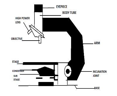
Compound Microscope Parts - Labeled Diagram and their Functions There are three major structural parts of a compound microscope. The head includes the upper part of the microscope, which houses the most critical optical components, and the eyepiece tube of the microscope. The base acts as the foundation of microscopes and houses the illuminator. The arm connects between the base and the head parts.
Label Microscope Diagram - EnchantedLearning.com Using the terms listed below, label the microscope diagram. arm - this attaches the eyepiece and body tube to the base. base - this supports the microscope. body tube - the tube that supports the eyepiece. coarse focus adjustment - a knob that makes large adjustments to the focus. diaphragm - an adjustable opening under the stage, allowing ...
Microscopy: Intro to microscopes & how they work (article) - Khan Academy Magnification is a measure of how much larger a microscope (or set of lenses within a microscope) causes an object to appear. For instance, the light microscopes typically used in high schools and colleges magnify up to about 400 times actual size. So, something that was 1 mm wide in real life would be 400 mm wide in the microscope image.
How To Draw A Microscope 🔬 - YouTube #microscope #howtodraw #adimushowThis is an easy and simple drawing of microscope .This will teach you how to draw and label a microscope .This is a step-by-...
Label a Microscope - Storyboard That Create a poster that labels the parts of a microscope and includes descriptions of what each part does. Click "Start Assignment". Use a landscape poster layout (large or small). Search for a diagram of a microscope. Using arrows and textables label each part of the microscope and describe its function. Copy This Storyboard.











![How To Draw A Microscope Step by Step - [12 Easy Phase]](https://easydrawings.net/wp-content/uploads/2021/01/Overview-for-Microscope-drawing.jpg)


Post a Comment for "42 draw and label microscope"