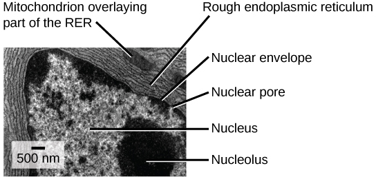
39 label the transmission electron micrograph of the mitochondrion
Label This Transmission Electron Micrograph / Microscopy Innovations ... Label the transmission electron micrograph of the nucleus. 0 nucleus rences mitochondrion heterochromatin peroxisome vesicle ular bumit . Scanning Transmission Electron Micrograph Stem Vesicles Were Download Scientific Diagram from Labeling nuclear proteins with electron dense probes in living cells. 0 nucleus rences ... Draw the structure of a mitochondrion as seen in an electron micrograph ... 411 6)a) Draw the structure of a mitochondrion as seen in an electron micrograph. [5] B) Describe the central role of acetyl (ethanoyl) CoA in carbohydrate & fat metabolism. [5] Acetyl CoA is formed in both carbohydrate and fat metabolism.
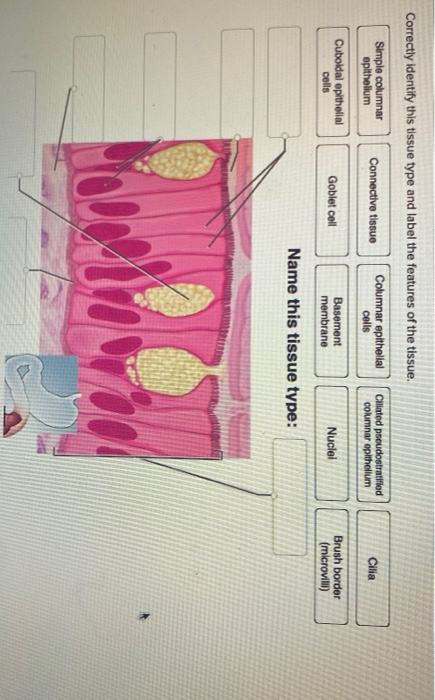
Solved Label the transmission electron micrograph of the - Chegg Transcribed image text: Label the transmission electron micrograph of the cell. 0 Nucleus rences Mitochondrion Heterochromatin Peroxisome Vesicle ULAR bumit Click and drag each label into the correct category to indicate whether it pertains to the cytoplasm or the plasma membrane.

Label the transmission electron micrograph of the mitochondrion
Bio101 - Ch 6 HW Flashcards | Quizlet Study with Quizlet and memorize flashcards containing terms like Which of the following choices correctly matches a tool and its proper application? See Concept 6.1 -cell fractionation to study the function of specific organelles -light microscopy to study the internal structure of cilia -transmission electron microscopy (TEM) to study the surfaces of preserved cells -transmission electron ... Transmission electron microscopy. (A) Endothelial cells of the control ... Lysosomal vesicles (arrow) were usually present in cultured cells. (B) Mitochondria (arrows) and endoplasmic reticulum with normal structure ( ϫ 12,000). ... Transmission electron microscopy. (A ... Transmission electron microscopy of iron oxide-labeled human ... We found that 70% of mitochondria are released from the hydrogel within 20 minutes at 37°C, that the respiratory capacity of hydrogel-released mitochondria over 60 minutes was greater than those ...
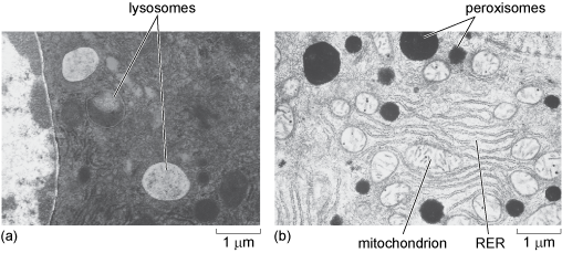
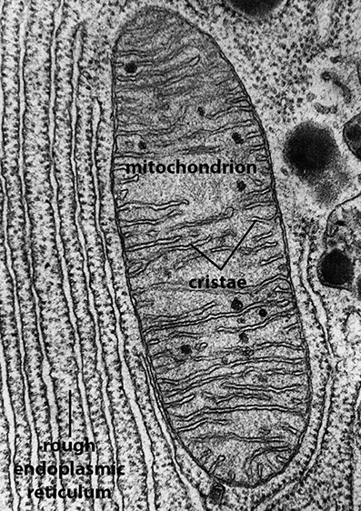
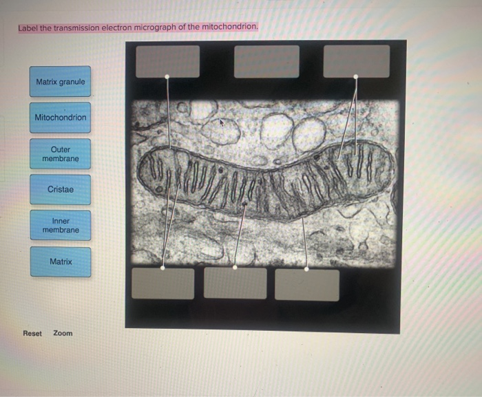
Label the transmission electron micrograph of the mitochondrion. Solved Label the transmission electron micrograph of the - Chegg Explanation - Mitochondrion is filamentous or globular in shape, occur in variable numbers from a few hundred to few thousands in different cells. It … View the full answer Transcribed image text: Label the transmission electron micrograph of the mitochondrion. Matrix granule Mitochondrion Outer membrane Cristae Inner membrane Matrix Reset Zoom AICE Biology Chapter 1: Plant Cell Electron Micrograph Labeling - Quizlet Start studying AICE Biology Chapter 1: Plant Cell Electron Micrograph Labeling. Learn vocabulary, terms, and more with flashcards, games, and other study tools. Transmission electron micrograph of mature MRCs with anti-Na+/K+-ATPase ... Download scientific diagram | Transmission electron micrograph of mature MRCs with anti-Na+/K+-ATPase immunogold labeling on tail of larvae adapted to 20 ppt at 5 dph. A: Mature MRC lying beneath ... Localization of A44 within mitochondria. Shown is transmission electron ... Download scientific diagram | Localization of A44 within mitochondria. Shown is transmission electron microscopy of sections of host cells either expressing A44-GFP (A) or incubated with exogenous ...
Uncertainty-Aware Label Rectification for Domain Adaptive Mitochondria ... To rectify noisy labels, a straightforward solution is to generate a label-wise mask m based on prediction C onfidence, i.e., m_C = \mathbbm {1} (p\ge \tau _p)+\mathbbm {1} (p< \tau _n), where \tau _p and \tau _n denote the thresholds for positive and negative labels, respectively. Generally, we have \tau _p\ge \tau _n. Solved Label the transmission electron micrograph based on - Chegg Question: Label the transmission electron micrograph based on the hints provided Mitochondrion Heterochromatin Plasma cell Nucleus Rough endoplasmic reticulum Nucleolus This problem has been solved! See the answer Show transcribed image text Expert Answer nucleus is the house of the genetic material which contains all the h … View the full answer Mitochondrial morphology and function: two for the price of one! This work represents a technical advance that allows the correlation of mitochondrial function and morphology with greater resolution and volume than has previously been feasible. LAY SUMMARY: Transmission electron microscopy (TEM) is a high-resolution technique used for the study of cells and their components, such as mitochondria. Coupling factor B affects the morphology of mitochondria - PMC I thank Dr. George Sachs for access to a laser scanning confocal microscope LSM 510, Dr. Olga Vagin for advice on confocal microscopy, Marianne Cilluffo for preparing specimens and for help with transmission electron microscopy, and Dr. William C. Claycomb for providing the HL-1 cells. This work was supported by NIH grant R01GM066085.
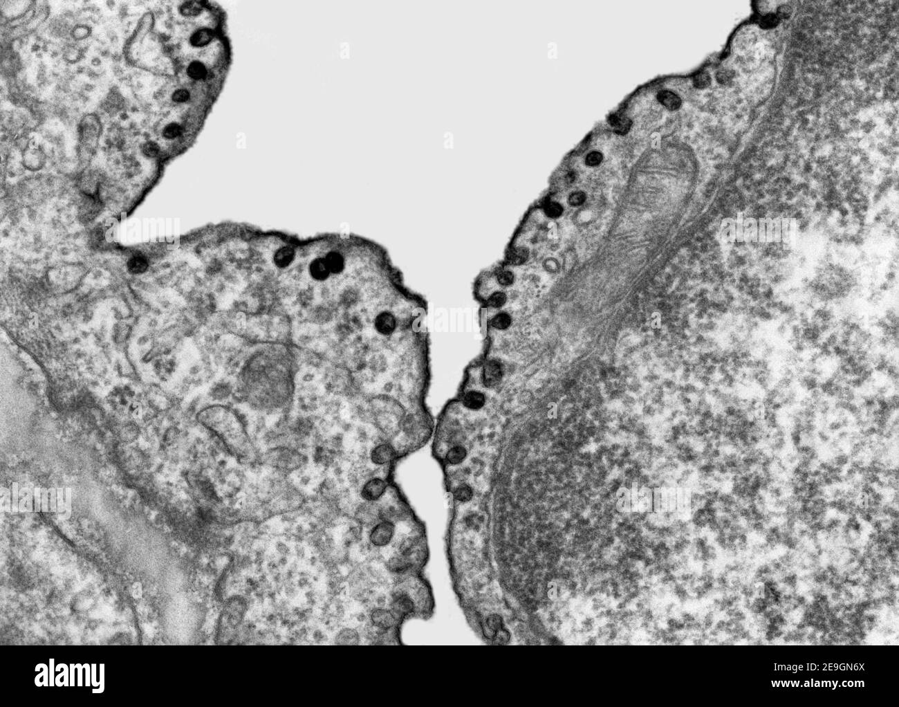
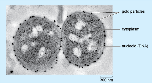
Transmission electron microscopy of C. parvum ... - ResearchGate Transmission electron microscopy of C. parvum sporozoites showing the relict mitochondrion, its relationship to other organelles, and Cp-mtHSP70-speci fi c immunogold localization within this or ... Electron Micrographs - University of Oklahoma Health Sciences Center Electron Micrographs Below is a collection of electron micrographs with labelled subcellular structures that you should be able to identify. Also, be sure to observe any electron micrographs which are made available in the laboratory by the instructor. Labeling the Cell Flashcards | Quizlet outside the cell wall Label the transmission electron micrograph of the cell. Label the transmission electron micrograph of the mitochondrion. Label the transmission electron micrograph of the nucleus. membrane bound organelles golgi apparatus, mitochondrion, lysosome, peroxisome, rough endoplasmic reticulum nonmembrane bound organelles Assessing mitochondria biogenesis - PubMed Mitochondria have their own DNA (mtDNA) and hence biogenesis of mitochondria requires a coordination of nuclear and mtDNA, both of which encode for mitochondria proteins. ... and measurement of mitochondria mass/volume in histological sections using fluorescent mitochondria dyes and light microscopy or transmission electron microscopy to yield ...
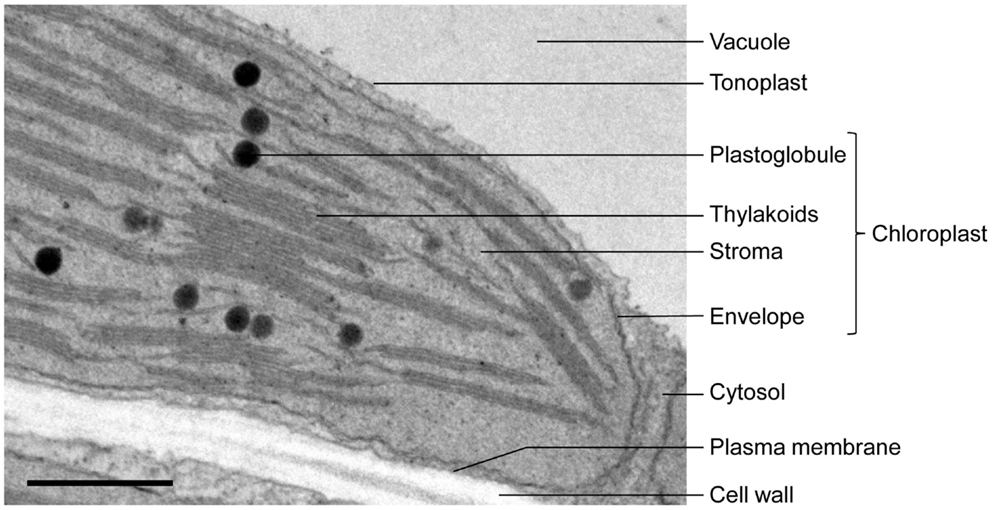
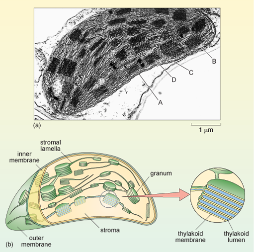
Chloroplast - Wikipedia
Transmission electron microscopy techniques - University of Otago The cytoplasm is full of mitochondria, lipid droplets, transparent vesicles, and an MII nucleus. Correlative TEM. Breast cancer cells by Sharon Lequeuex, read more below. Correlative microscopy involves using multiple microscope systems to observe the same specimen, most commonly light and transmission electron microscopy.
Transmission electron microscopy hi-res stock photography and ...
PDF Identifying Organelles from an Electron Micrograph - Ms JMO's Biology ... The electron micrograph displayed below illustrates many of the plant cell characteristics discussed The cell wall, large central vacuole and chloroplasts are clearly visible Also visible is the clearly defined nucleus containing chromatin
Solved Label the transmission electron micrograph of the ...
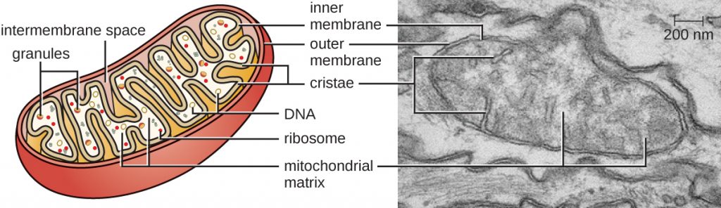
Microbiology Module 3 Flashcards | Quizlet The mitochondrion is the organelle involved in generating energy for the cell. It has a smooth continuous membrane surrounding its exterior. Inside, the inner membrane is folded into cristae. This inner membrane houses the proteins of the electron transport chain, involved in aerobic respiration. The fluid inside of the mitochondrion is called ...
Frontiers | When Proteomics Reveals Unsuspected Roles: The ...
Recent structural insight into mitochondria gained by microscopy diverse techniques such as high resolution scanning electron microscopy, transmission electron microscopy, electron microscope tomography and light microscopy have contributed a better understanding of mitochondrial compartmentalization, dynamic networks of mitochondria, intermembrane bridges, segregation of mitochondrial dna and contacts with …

Lewy pathology in Parkinson's disease consists of a crowded ...
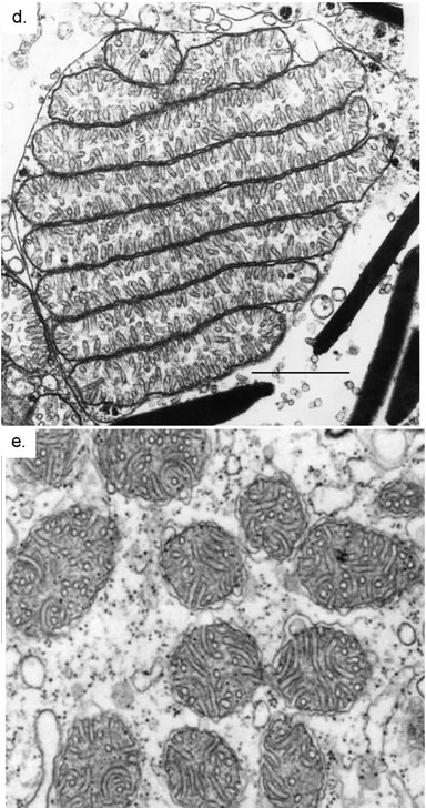
Electron Micrographs of Cell Organelles | Zoology - Biology Discussion It is an electron micrograph of cell's largest and most important organelle - the mitochondria and is characterized by the following features (Fig. 7 & 8): (1) The name mitochondria was given by Benda (1898) and their ma n function was brought to light by Kingsbury (1912).
Transmission Electron Micrograph (TEM) showing mitochondria ...
Mitochondria under the microscope — Science Learning Hub Transmission electron microscopy (left) shows the complex internal membrane structure of mitochondria, and electron tomography (right) gives a three-dimensional view. Microscopes have been crucial for our understanding of mitochondrial structure and function. Mitochondria are visible under the light microscope although little detail can be seen.
What is a diagram of a plant and animal cell under an ...
Transmission Electron Microscopy - Quizlet Examination of a cell by transmission electron microscopy reveals a high density of ribsomes in the cytoplasm. This observation suggests that this cell is actively producing large amounts of which molecules? ribosomes the nuclear lamina is an array of intermediate filaments that line the inner side of the nuclear membrane.

Cristae formation—linking ultrastructure and function of ...
Label the transmission electron micrograph of the nucleus. Label the transmission electron micrograph of the cell. 0 Nucleus rences Mitochondrion Heterochromatin Peroxisome Vesicle ULAR bumit Click and drag each label into the correct category to indicate whether it pertains to the cytoplasm or the plasma...
The mitochondrial compartment
Live-cell STED nanoscopy of mitochondrial cristae Dynamics of mitochondrial cristae. ( a) Transmission electron microscopy (TEM) of mitochondria from HeLa cells. ( b) Dual-color live-cell imaging of mitochondria. HeLa cells stably expressing...
Transmission electron micrographs of mitochondria, site of ...
Light and Electron Microscopy Study of Glycogen Synthase Kinase-3β in ... Examination by transmission electron microscopy revealed highly specific subcellular localization of GSK3β in neurons and astrocytes. At the subcellular level, GSK3β was present in the rough endoplasmic reticulum, free ribosomes, and mitochondria of neurons and astrocytes.
A tour of the cell: View as single page
Transmission electron microscopy of iron oxide-labeled human ... We found that 70% of mitochondria are released from the hydrogel within 20 minutes at 37°C, that the respiratory capacity of hydrogel-released mitochondria over 60 minutes was greater than those ...
Biology, The Cell, Cell Structure, The Endomembrane System ...
Transmission electron microscopy. (A) Endothelial cells of the control ... Lysosomal vesicles (arrow) were usually present in cultured cells. (B) Mitochondria (arrows) and endoplasmic reticulum with normal structure ( ϫ 12,000). ... Transmission electron microscopy. (A ...
Frontiers | Mitochondrial Morphology and Mitophagy in Heart ...
Bio101 - Ch 6 HW Flashcards | Quizlet Study with Quizlet and memorize flashcards containing terms like Which of the following choices correctly matches a tool and its proper application? See Concept 6.1 -cell fractionation to study the function of specific organelles -light microscopy to study the internal structure of cilia -transmission electron microscopy (TEM) to study the surfaces of preserved cells -transmission electron ...

Electron microscopy controls for mitochondria purified by ...

Transmission Electron Micrograph (TEM) showing mitochondria ...
What is a diagram of a plant and animal cell under an ...

Mitochondrial Structure-Function Correlation | Celebrate ...

Unique Characteristics of Eukaryotic Cells-Simplified ...

Solved Mitochondrion Nucleus Vesicle Peroxisome | Chegg.com

IB Biology Skills Practice Flashcards | Quizlet
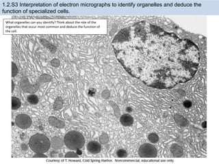
1.2 Obj Notes and Practice
A tour of the cell: View as single page

Mitochondrion - Wikipedia

Pin page

Cell Structures ‹ OpenCurriculum

Electron micrograph of a mitochondrion in a cell of the bat ...
A tour of the cell: View as single page

Dynamics of mitochondrial cristae. (a) Transmission electron ...
BIOL 230 Lecture Guide - Electron Micrograph of Mitochondria
Solved Label the transmission electron micrograph of the ...
3.4 Unique Characteristics of Eukaryotic Cells – Microbiology ...

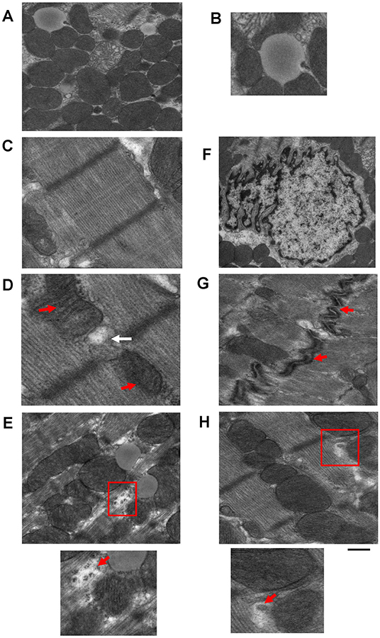
The morphology of mitochondria. (a) Thin-section electron ...

Mitochondrial morphology and function: two for the price of ...

An electron micrograph showing various orientations of ...

Mitochondria in cultivated fibroblasts visualized by ...

Labeling the Cell Flashcards | Quizlet
445 Transmission electron microscope Images, Stock Photos ...
9700 QR Dynamic Papers Biology al Cambridge

Transmission electron micrograph of mitochondria - Stock ...
Post a Comment for "39 label the transmission electron micrograph of the mitochondrion"